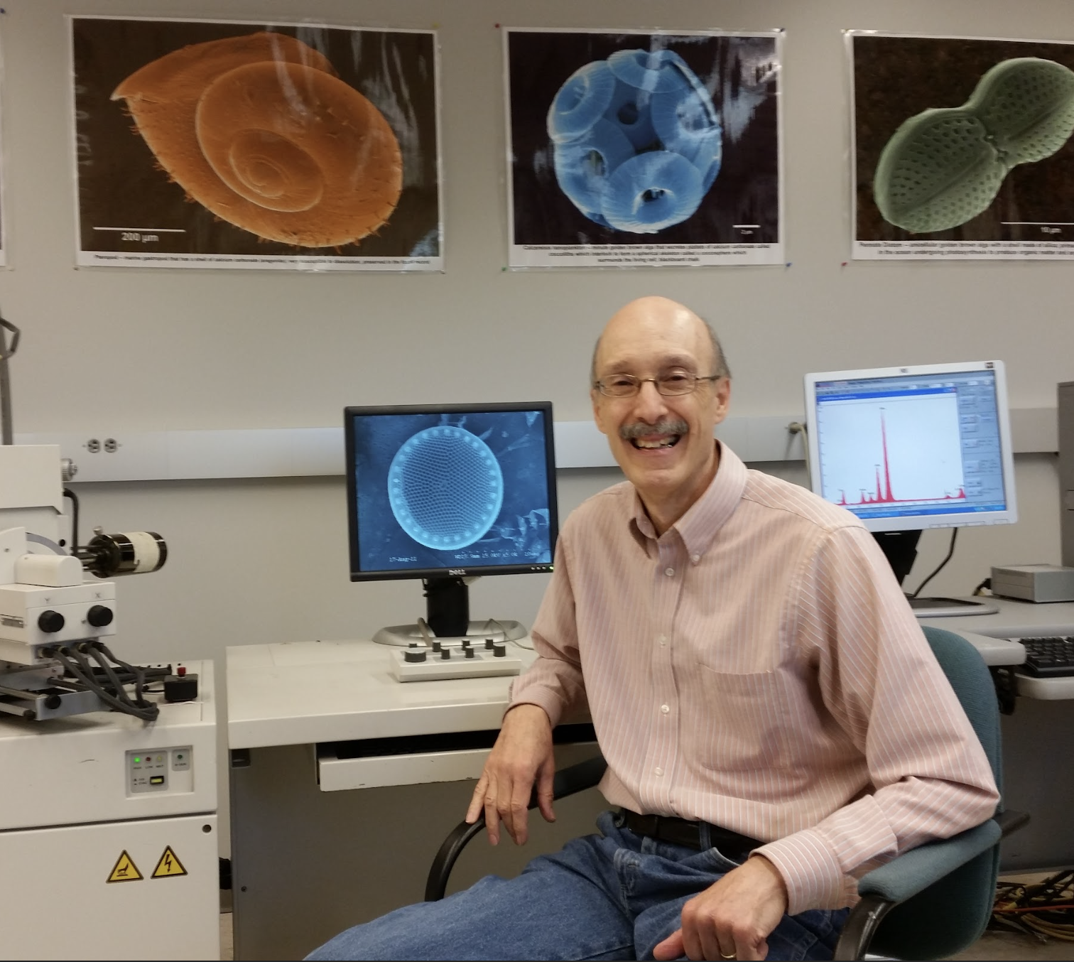
About Tony Greco
Education
M.S. in Biology Western Kentucky University, 1978
B.A. in Biology La Salle University, 1976
Work
Electron Microscope Manager at the College of Marine Science, University of South Florida 1980-2020
Managed the Transmission and Scanning Electron microscopes including specimen preparation equipment . Collaborated with faculty on numerous marine research projects. Taught undergraduate and graduate classes on the theory and use of the microscopes.
Lab technician at the University of Pennsylvania 1978-1980
Tony’s electron microscope images have been published in numerous scientific journals including the cover of Nature magazine.
You may contact Tony at oceanmicrocosm@gmail.com
How did you first become interested in microscopy?
I first became interested in science in general through my uncle who was a Professor of Biology at St. Bonaventure University in NY in the 70’s. He showed me fascinating images of plant sections using light microscopes in his lab. In graduate school at Western Kentucky University, I took some courses in transmission electron microscopy (TEM) and was stunned at the incredible cellular detail one could obtain. After that, I was hooked.
You were hired by the USF College of Marine Science in 1980, and according to your CV, made a move from Pennsylvania. Who was Dean then / Who hired you? What was the driving force behind the big move / what were you most excited about?
After graduating in 1978 with my Masters in biology, I spent a summer as an intern at the Department of Agriculture in the electron microscope lab in Philadelphia where I did research using the TEM on Phytophthora infestans, the fungus that caused the potato famine in Ireland in the 1840’s. I also learned how to use the scanning electron microscope (SEM). In the fall of that year, I landed a job at the University of Pennsylvania where I worked in the electron microscopy lab doing metallurgy research for 2 years. I was able to add x-ray microanalysis which allows elemental sample identification to my skill set. While I enjoyed my job at U of P, I was anxious to return to biological TEM and SEM and started looking for a new position.
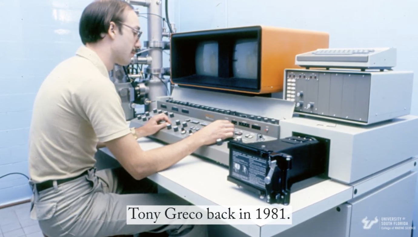
In 1980, Dr. John Steinmetz hired me to run a new SEM lab in the Department of Marine Science at USF so I packed up my world’s belongings and drove 1100 miles in my VW rabbit (no AC) to St. Petersburg, FL. It seemed like such an exciting new opportunity to run my own lab. John had just purchased a new SEM and showed me an old shower stall (MSL 233) where it would be housed. He immediately offered an SEM course where he taught the lectures and I ran the labs. This was before digital imaging so the students (all men) learned darkroom photography. When John left in 1982 for a job with the oil industry, I took over teaching both lecture and labs. The campus wasn’t very big in those days with just a few buildings. The entire library was housed in two rooms off the student lounge in MSL. The chair of the Dept of Marine Science (it wasn’t a college then so there was no Dean) at the time was Dr. Bill Sackett.
In 1994 an NSF grant and some seed money led to the purchase of a new Hitachi TEM which was installed in the new KRC building. The TEM was purchased mainly to obtain high resolution images of marine bacteria and viruses. I was then able to offer a graduate TEM course. Over the years I ‘ve used the TEM and SEM to look at everything from marine phytoplankton and zooplankton to bugs, cement and even micrometeorites in the lab.
What have you enjoyed most about your role as Electron Microscope Manager?
What I’ve most enjoyed most about my job has been working with all the incredibly talented faculty, staff and students I’ve collaborated with over the years on a wide variety of diverse research projects including discoveries of new viruses in copepods and seagrasses, the effects and recovery of benthic foraminifera from the Deepwater Horizon oil spill, and the characterization of Asian and Saharan dust particles.
What do you recall as your proudest microscopy moment?
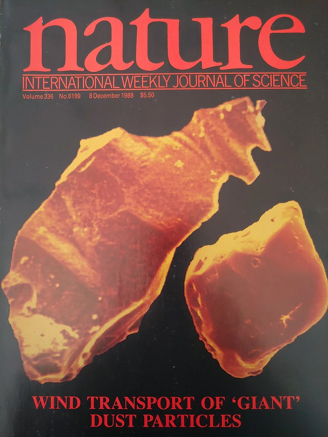
My proudest moment as a microscopist was making the cover of Nature magazine in December of 1988. Nature had just agreed to publish our article on the long range transport of large sand grains from dust storms in the Gobi desert when Peter Betzer suggested I submit a few of my SEM images of the sand grains as a potential cover photo. I was shocked when they selected my image. It also was a great honor to win the Costello Interdisciplinary Engineering award in 2017 which is a bit humorous as I’m not an engineer.
Lastly, one of the most significant things I’ve been involved with is chairing the Marine Science Safety Committee. Safety was not a huge priority in the 80’s and 90’s and I’m thrilled that the college now has regular lab inspections and faculty, staff and student lab safety training sessions.
Most memorable experience in the lab? 40 years is a wonderfully long time. Any favorite memory “snapshots” / memories come to mind (microscope-related or not)?
The community outreach events were very rewarding such as the St. Pete science festival, Oceanography Camp for Girls and the Great American Teach In as well as hosting numerous lab tours over the years. Most of the general public I find has no idea that the oceans are filled with bacteria and viruses. I always receive a big reaction to my 3-D stereo images and would routinely display some everyday samples like bugs to stimulate their interest. The recent science as art forum hosted by Mya Breitbart’s lab in January was very unique as several artists were inspired to make sculptures based on my SEM images. One artist even performed a dance with my SEM images scrolling on a giant screen.
Many of the memories over the years involved the interaction with my students. One particular grad student called me at home a little before midnight to ask if I could run down to the lab and change the filament on the SEM which had blown so she could finish her project. Another female student called me the night before a major exam tearfully begging me to reschedule it because her boyfriend had just broken up with her. Finally there was a struggling international student who kept bringing me home cooked food in a veiled attempt to bring up his grade. Unfortunately for him it didn’t work. Maybe my food standards are too high. I also enjoyed offering the SEM lecture and lab course to undergrads in the last 5 years. They had a real sense of wonder and awe at the incredible images they could obtain of samples they prepared themselves.
What about the CMS had you stay in your role for 40 years? What’s its greatest charm?
As to why I stayed at CMS for 40 years, I found that I really enjoyed the mix of teaching and research, the people I interacted with and the fact that I could work at my own pace in a relatively low stress job. Although I was offered a higher paying job in 1991 at a government research lab in the heart of New York city, I turned it down after deciding I didn’t want to give up teaching to write research papers for a living or raise a family there.
What will you NOT miss in your post-USF / post-microscope manager life?
Probably the least favorite part of my job is scrambling for funding to pay for servicing the microscopes and billing people for work on the instruments. This year, the 26 year old Hitachi TEM finally died so with severe budget cuts looming due to Covid and a $500,000 price tag for a new TEM, its probably a good time to retire.
What’s next? How do you plan to spend your time when not clocking it in at the CMS?
I’m looking forward to doing some travelling in retirement especially to the Carolinas where my two daughters and five grandchildren live. I’ll also be doing some volunteering in the community with my wife once the pandemic ends as well as having more time for a little golf and music. I’m taking bluegrass banjo lessons which has been immensely challenging.
Reprinted from Rising Tides Newsletter - December 2020, College of Marine Science University of South Florida